What Does a Cavity Look Like? Signs by Stage
What does a cavity look like? See real signs at every stage, from chalky white spots to dark holes, and when to see a dentist.
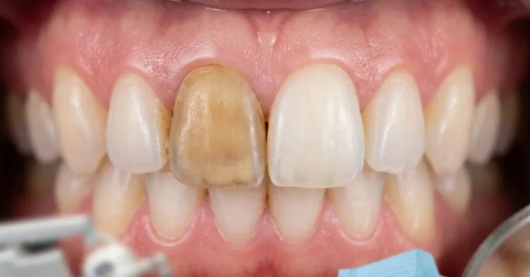
A cavity usually starts as a dull white or chalky spot on the tooth, then darkens to tan, brown, or black as the decay spreads, and eventually breaks the surface into a visible pit or hole. What it looks like at any given moment depends entirely on how far the decay has progressed, which is why two people can both have “a cavity” and see almost nothing alike in the mirror.
Nearly everyone deals with this at some point. The CDC reports that nine out of ten U.S. adults over 20 have had a cavity in a permanent tooth, and more than half of adolescents aged 12 to 19 have too. Knowing what to look for, and where cavities like to hide, is the difference between a small fluoride treatment and a root canal. This guide walks through every visible stage of a cavity, the spots that aren’t cavities at all, and exactly when a dark spot on your tooth means it’s time to call a dentist.
The 5 Stages of a Cavity, and What Each One Looks Like (Gap-fill)
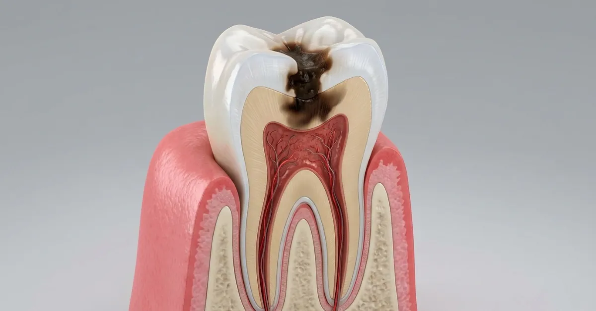
Tooth decay doesn’t jump straight from healthy enamel to a hole. Dentists generally describe it moving through five stages, and the visible signs change at each one.
| Stage | What it looks like | What’s happening | Reversible? |
|---|---|---|---|
| 1. Demineralization | Dull, chalky white spot, often near the gumline | Acid is pulling minerals out of the enamel before any structural damage occurs | Yes, with fluoride and better hygiene |
| 2. Enamel decay | White spot darkens to tan or light brown; surface may feel slightly rough | The enamel surface has broken down and a true cavity has formed | No, needs a filling |
| 3. Dentin decay | Spot deepens to dark brown or black; tooth may look slightly sunken | Decay has reached the softer dentin layer underneath, where it spreads faster | No, needs a filling or crown |
| 4. Pulp involvement | Visible dark hole; tooth may look discolored or gray | Bacteria have reached the nerve and blood vessels at the tooth’s core | No, usually needs a root canal |
| 5. Abscess | Swelling in the gum, sometimes a visible bump near the root | Infection has spread past the root tip into the surrounding bone | No, needs urgent dental treatment |
The only stage you can reverse at home is the first one. Once a white spot turns brown or a hole forms, the enamel has physically broken down, and only a dentist can restore it, not a toothpaste or a mouth rinse.
Stage 1: White Spots (Demineralization)
This is the earliest visible sign of a cavity, and the one people miss most often. Look for a dull, opaque white patch that doesn’t shine the way the rest of the tooth does, usually right along the gumline or in the grooves of a molar. A classic example is the white blotches that show up on teeth after braces come off, in the spots where plaque sat against the brackets for months.
At this stage there’s no pain, no hole, and often no sensitivity at all. That’s actually good news: demineralization can be stopped and even reversed with fluoride treatment, better brushing technique, and cutting back on frequent sugar exposure. If you catch a cavity here, you may never need a filling.
Stage 2: Enamel Decay
If the white spot isn’t treated, it starts to darken, first to a light tan, then to brown. This color change means the enamel surface has actually collapsed and a physical cavity, not just a weak spot, now exists. You might notice the area feels slightly sticky or rough if you run your tongue over it, though many people still feel nothing at all, since enamel itself has no nerve endings.
This is the point where the damage stops being reversible. A dentist will typically clean out the decayed area and place a filling to stop it from spreading further.
Stage 3: Dentin Decay
Once decay breaks through the enamel into the dentin underneath, things move faster. Dentin is softer than enamel and full of microscopic channels that lead straight to the nerve, so acid attacks the tooth much more quickly here than it did on the surface. Visually, the spot deepens to dark brown or black, and you may start to notice real symptoms: a sharp twinge when you eat something cold, sweet, or hot, especially if it lingers after the food or drink is gone.
Stage 4: Pulp Involvement
When decay reaches the pulp, the living center of the tooth that holds nerves and blood vessels, the cavity is usually an obvious dark hole you can see or feel with your tongue. Pain tends to become more constant at this stage rather than triggered only by hot or cold. A root canal is typically needed to remove the infected pulp and save the rest of the tooth structure.
Stage 5: Abscess
Left untreated, an infected pulp can lead to a tooth abscess, a pocket of pus that forms at the root tip after bacteria push past the tooth and into the surrounding bone. You may not be able to see the infection itself, but you’ll often see swelling in the gum near the tooth, sometimes with a small bump, along with intense pain and possibly a bad taste or fever. This is a dental emergency and needs same-day attention.
Cavity Colors: What White, Brown, and Black Spots Mean
Color is one of the fastest ways to gauge how advanced a cavity is, but it isn’t the whole story on its own.
- White spots: Almost always early demineralization. Dull and chalky, not shiny like healthy enamel. Most reversible stage.
- Yellow or light tan spots: Enamel has started to break down; a true cavity has likely formed underneath.
- Brown spots: Decay has been active for a while and has penetrated further into the tooth. Some brown spots are old, arrested decay that has hardened and stopped progressing, which only a dentist can confirm with an exam or X-ray.
- Black spots or black cavities: Usually the most advanced visible stage, meaning the decay has been present for a long time and a significant amount of tooth structure has broken down. Black cavities are more likely to need a crown or root canal rather than a simple filling.
- Orange or green staining: Less common, and in children especially, this can point to a specific bacterial infection rather than typical decay. It’s still worth a dental visit either way.
Darker generally means older and deeper, but color alone can’t tell you whether a spot is a stain, an old arrested lesion, or active decay. That distinction takes a dental exam.
Cavity or Just a Stain? How to Tell the Difference (Gap-fill)
This is one of the most common points of confusion, and most cavity guides skip right past it. A cavity and a surface stain can look similar at a glance, but a few details usually separate them:
- Texture: Stains sit on a smooth surface. A cavity often feels rough, sticky, or slightly soft when you run your tongue over it or when a dentist tests it with an explorer tool.
- Shape: Stains tend to follow the natural contour of the tooth or coat a wide, even area. Cavities are more likely to be localized to a pit, groove, or the edge of a filling.
- Sensation: A stain causes no sensitivity. A cavity, once it reaches the dentin, often responds to cold, heat, or sweets, sometimes with lingering discomfort after the stimulus is gone.
- Cause: Surface stains commonly come from coffee, tea, red wine, tobacco, or certain foods, and they wipe or polish away more easily during a cleaning. Cavity discoloration comes from mineral loss and bacterial acid, and it won’t buff out.
- Confirmation: Only a dentist can tell the two apart with certainty, usually through a visual and tactile exam plus a bitewing X-ray, since some early decay looks nearly identical to a stain to the naked eye.
If a spot doesn’t come off with a professional cleaning and the tooth feels different than the surrounding enamel, treat it as a possible cavity until a dentist says otherwise.
Where Cavities Show Up First
Cavities don’t form randomly. They cluster in the spots where plaque sits undisturbed the longest, which is also why some of the earliest ones are the hardest to spot in a mirror.
- Chewing surfaces of molars and premolars: The pits and grooves on the top of your back teeth trap food and plaque even with regular brushing, and this is where a large share of childhood cavities start.
- Between teeth: Floss-only territory. Interdental cavities often don’t show up until a dentist takes a bitewing X-ray, since you can’t see between the teeth in a mirror.
- Along the gumline: Plaque that isn’t brushed away at the gum margin can demineralize the enamel right at that border, showing up as a white or brown band low on the tooth.
- Root surfaces: In adults with gum recession, the exposed root surface has no enamel covering it at all, so decay here moves faster and is common in older adults.
- Around old fillings or crowns: The edge where a filling meets natural tooth is a frequent site for new, smaller decay, sometimes called recurrent decay.
What a Cavity Looks Like on a Dental X-Ray
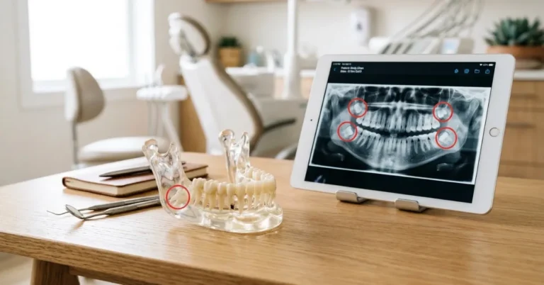
Not every cavity is visible to the eye, and X-rays catch decay long before it would ever show up in a mirror. On a bitewing X-ray, healthy enamel and dentin appear as light, solid areas, while decay shows up as a darker gray or black shadow, since decayed tooth structure is less dense and lets more radiation pass through.
Small cavities, especially the kind forming between teeth, are frequently invisible on an X-ray in their earliest stage simply because they aren’t yet large enough to change the tooth’s density in a detectable way. This is one reason dentists recommend routine checkups on a schedule rather than waiting for visible symptoms: a cavity can be developing between two teeth for months before it’s large enough to see on film or feel with a fork.
Symptoms That Go Along With Visual Signs
A cavity’s appearance rarely tells the whole story by itself. These physical symptoms often show up alongside or before you notice a visible change:
- Sharp pain when biting down or chewing
- Sensitivity to hot, cold, or sweet foods and drinks, sometimes lingering after the trigger is gone
- A rough, jagged, or soft feeling when you run your tongue over the tooth
- Persistent bad breath or an unpleasant taste that doesn’t go away with brushing
- Visible pitting or a hole you can feel with your tongue or fingernail
- Food consistently getting stuck in the same spot
Early demineralization typically causes no symptoms at all, which is exactly why regular checkups matter more than waiting to feel something.
What Cavities Look Like in Children’s Teeth (Gap-fill)
Cavities in baby teeth and newly erupted permanent teeth often look slightly different than decay in adult teeth, and parents commonly miss them for that reason.
- Baby bottle tooth decay: Chalky white lines or bands near the gumline on the upper front teeth, often from a bottle or sippy cup used at bedtime with milk, formula, or juice. This is usually the first visible sign in toddlers.
- Faster progression: Baby teeth have thinner enamel and larger pulp chambers relative to their size, so decay can move from a white spot to a deep cavity faster than it would in an adult tooth.
- Molars are the most common site: The deep grooves on newly erupted six-year molars are a frequent starting point, and sealants are often recommended specifically to protect this area.
- Kids may not report pain: Younger children often can’t describe tooth sensitivity clearly, so a visible spot, a change in eating habits, or a child favoring one side of the mouth can be the only early clue.
The American Academy of Pediatric Dentistry recommends a child’s first dental visit by their first birthday or within six months of the first tooth erupting, specifically so early decay can be caught before it’s visible to a parent.
When to See a Dentist
Any visible spot, discoloration, or hole is worth a dental visit, even without pain, since the earliest and most reversible stage of a cavity causes no symptoms at all. See a dentist sooner rather than later if you notice:
- A white, brown, or black spot that wasn’t there before
- A tooth that feels rough, sticky, or has a visible pit
- Sensitivity to hot, cold, or sweets that lingers for more than a few seconds
- Pain when biting or chewing
- Swelling, a bump on the gum, or a bad taste that won’t go away, which can signal an abscess and needs urgent care
Waiting rarely makes a cavity smaller. A spot that would need a simple filling today can require a root canal or extraction a year from now if the decay is left to spread.
How to Prevent Cavities From Forming
Since plaque acid is the root cause at every stage, the prevention list stays fairly short and consistent:
- Brush twice a day for two minutes with fluoride toothpaste, angling the brush toward the gumline
- Floss once a day to clean the surfaces between teeth a toothbrush can’t reach
- Limit frequent snacking and sipping on sugary or acidic drinks, since it’s the frequency of acid exposure that matters more than the total amount
- Ask about fluoride treatments or dental sealants, especially for children’s molars
- Keep regular dental checkups and cleanings, even when nothing hurts, since X-rays catch decay long before it’s visible
Frequently Asked Questions
Can a cavity go away on its own?
Only at the very first stage, called demineralization, when it’s still a white spot with no hole. At that point, fluoride treatment and better oral hygiene can remineralize the enamel. Once the surface has broken down into a true cavity, it will not heal on its own and needs a dentist to treat it.
What does the beginning of a cavity look like?
The earliest visible sign is usually a dull, chalky white spot on the tooth surface, often near the gumline or in the grooves of a molar. It looks different from healthy enamel because it’s matte rather than glossy, but there’s typically no pain or hole yet.
Do all cavities turn black?
No. A cavity’s color depends on how long it’s been progressing. Many cavities are caught and filled while they’re still white or light brown. Black usually indicates decay that has been active for a longer period, though a dark spot can also be arrested decay that has stopped progressing, which only a dental exam can confirm.
Can you see a cavity between your teeth?
Sometimes, but not reliably. Interdental cavities are one of the most commonly missed types because they’re hidden from view in a mirror. Dentists typically catch these with bitewing X-rays during routine checkups, well before they’d be visible or cause pain.
Is a cavity painful?
Not always, and not at every stage. Early demineralization and even early enamel decay often cause no pain at all. Sensitivity to hot, cold, or sweets usually starts once decay reaches the dentin layer, and sharp or constant pain typically signals the decay has reached the pulp.
How fast does a cavity grow?
It varies widely based on diet, oral hygiene, saliva, and the tooth’s location, but a cavity in enamel can take anywhere from several months to a couple of years to reach the dentin. Once decay reaches the softer dentin layer, it tends to spread noticeably faster than it did in the harder enamel.
Key Takeaways
A cavity’s appearance changes as it progresses: dull white spots come first and are still reversible, then tan or brown discoloration as the enamel breaks down, then dark brown or black spots with visible pitting once decay reaches the dentin and beyond. Color and texture together, not just color alone, are what separate a cavity from an ordinary stain, and some of the most common cavities, the ones between teeth, aren’t visible in a mirror at all.
The most useful habit isn’t trying to self-diagnose every spot you find. It’s treating any new discoloration, rough patch, or lingering sensitivity as a reason to book a dental exam, since the earliest stage is also the only one you can still reverse without a filling.